
Darkfield Studies
Written by Ingrid Naiman
Wednesday, 28 February 2007
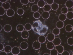
Darkfield Image of Normal Blood
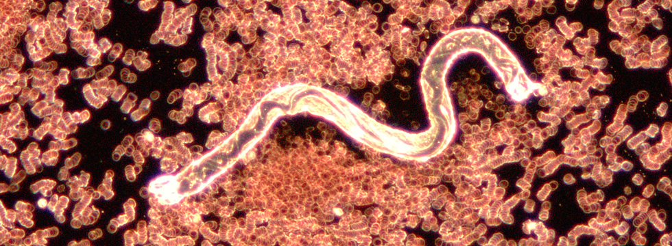
Darkfield microscopy is a special form of microscopy in which the light beam is split in such a way that the edges of objects in the samples are illuminated so that they appear as silhouettes against a dark background — as opposed to brightfield microscopy which allows the examination of specimens against an illuminated field — and which washes out the tiny and faint objects that can be seen only in darkfield.
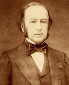
Claude Bernard
Historically, this form of microscopy has most often been associated with two critical ideas, usually attributed to Claude Bernard (1813-1878) and Antoine Béchamp, (1816-1908): that the milieu determines the risk of pathogenicity; and pleomorphism, the belief or doctrine that bacteria have multiple forms (depending on the milieu). Bernard held the first chair in physiology at the Sorbonne and was highly regarded as a true scientist, emphasizing the need to accept facts, even if they are opposed by prevailing opinions.
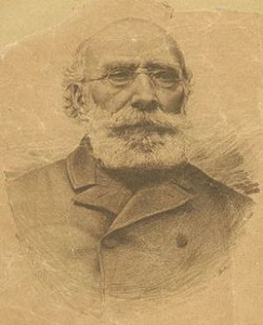
Antoine Béchamp
The insights of these illustrious Frenchmen influenced the work of Prof. Gunther Enderlein, Royal Raymond Rife, Wilhelm Reich, Gaston Naessens, Virginia Livingston-Wheeler, Dr. Alan Cantwell, and Prof. Erik Enby.
The method of observation is just that: a technological modification of a basic light microscope that allows one to view live samples as well as objects that are usually obscured by the bright
Privacy Policy || Report Technical Problems || Permission to Quote || Job Opportunities
Copyright Dr. Ingrid Naiman @ 2014-2020. All Rights Reserved.
For permission to quote, please contact the Institute for Invisible Epidemics.
Poulsbo, Washington, USA.